Vertebral Compression Fractures
Vertebral Compression Fractures
Incidence and Prevalence
Vertebral compression fractures (VCFs) are the most common fracture in patients with osteoporosis, affecting about 750,000 people annually. VCFs affect an estimated 25 percent of all postmenopausal women in the United States. The prevalence of this condition steadily increases as people age, with an estimated 40 percent of women age 80 and older affected. Although far more common in women, VCFs are also a major health concern for older men.
People who have sustained one osteoporotic VCF are at five times the risk of sustaining a second VCF. Occasionally a VCF can be present with either minor symptoms or no symptoms, but the risk still exists for additional VCFs to occur.
Causes
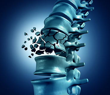
VCFs occur when the bony block or vertebral body in the spine collapses, which can lead to severe pain, deformity and loss of height. These fractures more commonly occur in the thoracic spine (the middle portion of the spine), especially in the lower part. While osteoporosis is the most common cause, these fractures may also be caused by trauma or metastatic tumors.
In people with severe osteoporosis, a VCF may be caused by simple daily activities, such as stepping out of the shower, sneezing vigorously or lifting a light object. In people with moderate osteoporosis, it usually takes increased force or trauma, such as falling down or attempting to lift a heavy object to cause a VCF. People with healthy spines most commonly suffer a VCF through severe trauma, such as a car accident, sports injury or a hard fall.
Metastatic tumors should be considered as the cause in patients younger than 55 with no history of trauma or only minimal trauma. The bones of the spine are a common place for many types of cancers to spread. The cancer may cause destruction of part of the vertebra, weakening the bone until it collapses.
Symptoms
The main clinical symptoms of VCFs may include any of the following, alone or in combination:
- Sudden onset of back pain
- An increase of pain intensity while standing or walking
- A decrease in pain intensity while lying on the back
- Limited spinal mobility
- Eventual height loss
- Eventual deformity and disability
Complications related to VCFs include:
- Segmental Instability
- Kyphosis
- Neurological Complications
Segmental Instability
When a fracture leads to a vertebral body collapse of more than 50 percent, there is a risk of segmental instability. The spinal segments work together to enable weight bearing, movement, and support of the entire spine. When one segment deteriorates or collapses to the point of instability, it can produce pain and impair daily activities. The instability ultimately results in quicker degeneration of the spine in the affected area.
Kyphosis
Kyphosis is a common disorder in older women who have osteoporosis and frequent VCFs. The front of the vertebrae will collapse and “wedge” due to the lack of normal vertebral space. Kyphosis leads to a more rounded thoracic spine. This deformity is sometimes referred to as hunchback or dowager’s hump.
Severe kyphosis may cause extreme and debilitating pain. The hunchback deformity may eventually compress the heart, lungs, and intestines. This in turn can lead to fatigue, shortness of breath, and loss of appetite.
Neurological Complications
If the fracture causes part of the vertebral body to place pressure on the spinal cord, the nerves and spinal cord can be affected. The normal space between the spinal cord and beginning of the spinal canal can be reduced if pieces of the broken vertebral body push into the spinal canal.
The narrowing of the spinal canal due to a VCF can lead to immediate injury to the spinal nerves, or can cause problems later from irritation of the nerves. The lack of space can also lower the supply of blood and oxygen to the spinal cord. This can lead to numbness and pain in the nerves that are affected. The nerves may lose some of their mobility when the space around them decreases, which can lead to nerve irritation and inflammation.
Diagnosis
While a diagnosis can usually be made through history and a physical examination, plain x-rays, computed tomography or magnetic resonance imaging, can help in confirming diagnosis, predicting prognosis, and determining the best treatment option for the patient.
- X-ray: Application of radiation to produce a film or picture of a part of the body can show the structure of the vertebrae and the outline of the joints. It will also show bone alignment, disc degeneration, and bony spurs which may irritate nerve roots.
- Computed tomography scan (CT or CAT scan): A diagnostic image created after a computer reads x-rays; can show the shape and size of the spinal canal, its contents, and the structures around it. This test may be performed in conjunction with a myelogram of the spine to provide additional information. This diagnostic study is ideal for showing bone detail including stenosis.
- Magnetic resonance imaging (MRI) : A diagnostic test that produces three-dimensional images of body structures using powerful magnets and computer technology; can show the spinal cord, nerve roots, and surrounding areas, as well as enlargement, degeneration, and tumors.
- Dual-energy x-ray absorptiometry (DXA or DEXA) or bone densitometry : This test is the established standard for measuring bone mineral density and can determine if osteoporosis exists. The scanner painlessly and rapidly directs x-ray energy from two different sources towards the bone being examined in an alternating fashion at a set frequency. A DEXA scan can detect small changes in bone mass and is also more flexible since it can be used to examine both the spine and the extremities. A scan of the spine, hip or the entire body requires less than four minutes.
Nonsurgical Treatment
Traditionally, people with severe pain from VCFs have been treated with bed rest, medications, bracing, or invasive spinal surgery, often with limited effectiveness. Pain secondary to acute vertebral fracture appears to be caused in part by vertebral instability (nonunion or slow-forming union) at the fracture site. VCF-related pain that is allowed to heal naturally can last as long as three months. However, the pain usually decreases significantly in a matter of days or weeks.
Bed rest may be advised for a short period of time, followed by a limitation on some activities. However, prolonged inactivity should be avoided.
Over-the-counter pain medications are often effective in relieving pain. Both acetaminophen and nonsteroidal anti-inflammatory drugs (NSAIDs) are commonly recommended. Narcotic pain medications and muscle relaxants are often prescribed, but only for short periods of time, due to the risk of addiction.
Back bracing can provide external support to limit the motion of fractured vertebrae, similar to the support a cast provides on a leg fracture. The rigid style of back brace limits spine-related motion greatly, which may help reduce pain.
While immediate treatment is essential to alleviating the pain and risks of the fracture, prevention of subsequent fractures is very important. Your physician may prescribe bone-strengthening drugs known as bisphosphonates (i.e.: Actonel, Boniva, and Fosamax) to help stabilize or restore bone loss.
When conservative treatment options have proven ineffective, two minimally invasive procedures, called vertebroplasty and kyphoplasty may be considered as treatment options. Recent advances in spinal procedures have reduced the need for invasive surgery, in many cases.
Vertebroplasty and Kyphoplasty
Vertebroplasty for the treatment of VCFs was introduced in the United States in the early 1990s. The procedure is usually done on an outpatient basis, although some patients stay in the hospital overnight. Vertebroplasty takes from one to two hours to perform, depending on the number of vertebrae being treated. The procedure may be performed with a local anesthetic and intravenous sedation or general anesthesia. Using x-ray guidance, a small needle containing specially formulated acrylic bone cement is injected into the collapsed vertebra. The cement hardens within minutes, strengthening and stabilizing the fractured vertebra. Most experts believe that pain relief is achieved through mechanical support and stability provided by the bone cement.
A newer procedure, called kyphoplasty, involves an added procedure performed before the cement is injected into the vertebra. First, two small incisions are made and a probe is placed into the vertebral space where the fracture is located. The bone is drilled and one balloon (called a bone tamp) is inserted on each side. The two balloons are then inflated with contrast medium (which are visualized using image guidance x-rays) until they expand to the desired height and removed. The spaces created by the balloons are then filled with the cement. Kyphoplasty has the added benefit of restoring height to the spine.
Patients with the following criteria may be considered candidates for vertebroplasty or kyphoplasty:
- Osteoporotic VCFs in any area of the spine that have been present for more than two weeks, causing moderate to severe pain, and unresponsive to conservative therapy
- Painful metastases and multiple myelomas
- Painful vertebral hemangiomas (benign, malformed vascular tumors composed of newly formed blood vessels)
- Vertebral osteonecrosis (a condition resulting from poor blood supply to an area of bone, which causes bone death)
- Reinforcement of a pathologically weak vertebral body before a surgical stabilization procedure
Patients with any of the following criteria should not undergo these procedures:
- A VCF that is completely healed or is responding effectively to conservative therapy
- A VCF that has been present for more than one year
- Greater than 80 to 90 percent collapse of the vertebral body
- Spinal curvature such as scoliosis or kyphosis that is due to causes other than osteoporosis
- Spinal stenosis or herniated discs with nerve or spinal cord compression and loss of neurological function not associated with a VCF
- Untreated coagulopathy (a disease or condition affecting the blood’s ability to coagulate)
- Osteomyelitis (an inflammation of the bone and bone marrow, usually caused by bacterial infection)
- Discitis (nonbacterial inflammation of an intervertebral disc or disc space)
- Significant compromise of the spinal canal caused by impeding bone fragment or tumor
Complication rates for vertebroplasty and kyphoplasty have been estimated at less than 2 percent for osteoporotic VCFs and up to 10 percent for malignant tumor-related VCFs. The benefits of surgery should always be weighed carefully against its risks. Although a large percentage of patients report significant pain relief after these two procedures, there is no guarantee that surgery will help every individual.