Cervical Spine
Cervical Spine
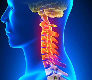
Your neck is part of a long flexible column, known as the spinal column or backbone, which extends through most of your upper body. The cervical spine (neck region) consists of seven blocks of bones (C1-C7 vertebrae), which are separated from one another by intervertebral discs. These discs allow the spine to move freely and act as shock absorbers during activity.
Attached to the back of each vertebral body is an arch of bone that forms a continuous hollow longitudinal space, which runs the whole length of your back. This space, called the spinal canal, is the area through which the spinal cord and nerve bundles pass. The spinal cord is bathed in cerebrospinal fluid (CSF) and surrounded by three protective layers called the meninges (dura, arachnoid, and pia mater).
At each vertebral level, a pair of spinal nerves exit through small openings called foramina (one to the left and one to the right). These nerves supply the muscles, skin and tissues of the body and thus provide sensation and movement to all parts of the body. The delicate spinal cord and nerves are further supported by strong muscles and ligaments that are attached to the vertebrae.
Cervical disc disease:
You may have been referred to a neurospecialist because of pain in your neck or shoulder, or tingling and numbness in your arms. You may also have experienced some weakness in your arms or hands.
Neck pain may be caused by disc degeneration, sprain of muscles or ligaments, narrowing of the spinal canal, arthritis, and, in rare cases, cancer or meningitis. You should consult a neurosurgeon for neck pain if:
- It occurs after an injury or blow to the head
- Fever or headache accompanies the neck pain
- Stiff neck prevents you from touching your chin to your chest
- Pain shoots down one arm
- There is tingling, numbness or weakness in your arms or hands
- Neck symptoms associated with leg weakness or loss of coordination in arms or legs.
- Your pain does not respond to over-the-counter pain medication
- Pain does not improve after a week
Age, injury, poor posture, or diseases such as arthritis can lead to degeneration of the bones or joints of the cervical spine, causing disc herniation or bone spurs to form. Sudden severe injury to the neck may also contribute to disc herniation, whiplash, blood vessel destruction, vertebral injury, and, in extreme cases, permanent paralysis. Herniated discs or bone spurs may cause a narrowing of the spinal canal or the small openings through which spinal nerve roots exit.
Pressure on the spinal cord in the cervical region can be a very serious problem because virtually all of the nerves to the rest of the body have to pass through the neck to reach their final destination (arms, chest, abdomen, legs). This can potentially compromise the function of many important organs.
Cervical stenosis:
Cervical stenosis occurs when the spinal canal narrows and compresses the spinal cord and is most frequently caused by aging. The discs in the spine that separate and cushion vertebrae may dry out. As a result, the space between the vertebrae shrinks, and the discs lose their ability to act as shock absorbers. At the same time, the bones and ligaments that make up the spine become less pliable and thicken. These changes result in a narrowing of the spinal canal. In addition, the degenerative changes associated with cervical stenosis can affect the vertebrae by contributing to the growth of bone spurs that compress the nerve roots. Mild stenosis can be treated conservatively for extended periods of time as long as the symptoms are restricted to neck pain. Severe stenosis requires referral to a neurosurgeon.
Symptoms:
- Neck or arm pain
- Numbness and weakness in both hands
- Unsteady gait when walking
- Muscle spasms in the legs
- Loss of coordination
Diagnosis:
Diagnosis is made by a neuro doctor based on your history, symptoms, a physical examination, and results of tests, including the following:
- Computed tomography scan (CT or CAT scan): A diagnostic image created after a computer reads x-rays; can show the shape and size of the spinal canal, its contents, and the structures around it.
- Electromyogram and Nerve Conduction Studies (EMG/NCS): These tests measure the electrical impulse along nerve roots, peripheral nerves, and muscle tissue. This will indicate whether there is ongoing nerve damage, if the nerves are in a state of healing from a past injury, or whether there is another site of nerve compression.
- Magnetic resonance imaging (MRI): A diagnostic test that produces three-dimensional images of body structures using powerful magnets and computer technology; can show the spinal cord, nerve roots, and surrounding areas, as well as enlargement, degeneration, and tumors.
- Myleogram: An x-ray of the spinal canal following injection of a contrast material into the surrounding cerebrospinal fluid spaces; can show pressure on the spinal cord or nerves due to herniated discs, bone spurs or tumors.
- X-ray: Application of radiation to produce a film or picture of a part of the body can show the structure of the vertebrae and the outline of the joints.
Treatment:
Nonsurgical treatment is the first approach in patients with common neck pain not involving trauma. For example, many patients with cervical disc herniations improve with conservative treatment and time and do not require surgery. Conservative treatment includes pain medication, bed rest, reduction of physical activity, and physiotherapy. Your doctor may prescribe medications to reduce the pain or inflammation and muscle relaxants to allow time for healing to occur. An injection of corticosteroids may be used to temporarily relieve pain.
Surgery:
You may be a candidate for surgery if:
- Conservative therapy is not helping
- You experience progressive neurological symptoms involving your arms and legs
- You experience difficulty with balance or walking
- You are in otherwise good health
There are several different surgical procedures which can be utilized, the choice of which is influenced by the severity of your case. In a small percentage of patients, spinal instability may require that spinal fusion be performed, a decision that is generally determined prior to surgery. Spinal fusion is an operation that creates a solid union between two or more vertebrae. Various devices (like screws or plates) may be used to enhance fusion and support unstable areas of the cervical spine. This procedure may assist in strengthening and stabilizing the spine and may thereby help to alleviate severe and chronic neck pain.
Anterior Cervical Discectomy
This operation is performed on the neck to relieve pressure on one or more nerve roots, or on the spinal cord. The cervical spine is reached through a small incision in the anterior (front) of your neck. If only one disc is to be removed, it will typically be a small horizontal incision in the crease of the skin. The space left between the vertebrae may be left open or filled with a small piece of bone through spinal fusion. In time, the vertebrae may fuse or join together.
Anterior Cervical Corpectomy
This operation is performed in conjunction with the anterior cervical disectomy. The corpectomy is often done for multi-level cervical stenosis with spinal cord compression caused by bone spur formations. In this procedure, the neurosurgeon removes a part of the vertebral body to relieve pressure on the spinal cord. The space between the vertebrae is filled using a small piece of bone through spinal fusion. Your surgeon may select to use a metal plate that is screwed into the front of the vertebra to help the healing process.
Posterior Cervical Laminectomy
This procedure requires a small incision in the middle of your neck to remove bone spur formations or disc material. The foramen, the passage in the vertebrae of the spine through which the spinal nerve roots travel is enlarged, to allow the nerves to pass through.
Your neurosurgeon will remove a section of the lamina (the back bony part of the vertebrae) and ligament to find the exact area of the compression. An operating microscope is used to create an opening, and part of the lamina is removed to take pressure off the nerves and spinal cord. If needed, bone spurs, tissue and any disc fragments causing the compression are also removed.
Risks and Outcome:
Although complications are fairly rare, as with any surgery. The benefits of surgery should always be weighed carefully against its risks. Although a large percentage of cervical spine patients report significant pain relief after surgery.
Post-surgery:
Your doctor will give you specific instructions postsurgery and usually prescribe pain medication. Your doctor will help determine when you can resume normal activities such as returning to work, driving and exercising. Some patients may benefit from supervised rehabilitation or physical therapy after surgery. Discomfort is expected while you gradually return to normal activity, but pain is a warning signal that you might need to slow down.