Stereotactic Radiosurgery
Stereotactic Radiosurgery
Stereotactic radiosurgery uses sophisticated 3-D computerized imaging to precisely target a narrow X-ray beam and deliver a highly concentrated dose of radiation to the affected area. Stereotactic radiosurgery is not surgery in the conventional sense because there is no incision involved, and general anesthesia is not required for adults.
Staged radiosurgery, also known as fractionated stereotactic radiosurgery (FSR), is a process in which the total dose of stereotactic radiation is divided into several smaller doses of radiation, on separate days of treatment. Typically, this consists of two to five treatments. In some cases, staged treatment effectively kills the tumor while seemingly decreasing potential side effects compared with single-dose radiosurgery.
The radiation oncologist and neurosurgeon oversee treatment and interpret the results of these procedures.
Treatment Uses
Unlike conventional whole-brain radiation in which a person receives a small amount of radiation every day over several weeks just once in their lifetime, stereotactic radiosurgery may be repeated. Patients can receive stereotactic radiosurgery in addition to whole-brain radiation. This technology allows high doses of radiation to be delivered to the tumor with minimal exposure to surrounding healthy tissue.
Stereotactic radiosurgery is a noninvasive treatment option for many patients with abnormal blood vessels in the brain such as arteriovenous malformations, arteriovenous fistulas, brain tumors and trigeminal neuralgia.
Arteriovenous Malformations and Arteriovenous Fistulas
An arteriovenous malformation (AVM) is a tangle of abnormal and poorly formed blood vessels (arteries and veins) with a higher rate of bleeding than normal vessels. AVMs can occur anywhere in the body, but brain AVMs present substantial risks when they bleed. Dural AVMs occur in the covering of the brain, and are an acquired disorder that may be triggered by an injury.
An arteriovenous fistula (AVF) is an abnormal channel or passage between an artery and a vein that causes a disruption of normal blood flow patterns. A person may be born with a congenital AVF or may develop an acquired fistula after birth, often caused by an injury that damages the arteries.
Stereotactic radiosurgery targets concentrated radiation to the malformed vessels of the brain, causing the abnormal vessels to close off. Unfortunately, this treatment is usually limited to lesions less than 3.5 cm in diameter, and it may take up to two years to completely obliterate the lesion.
Intracranial Tumors
Stereotactic radiosurgery may be a treatment option for select patients with some metastatic brain tumors that arise from other parts of the body (such as the lungs) and for such primary brain tumors as the following:
- anaplatic astrocytomas
- chordomas
- craniopharyngiomas
- glioblastomas
- gliomas
- hemangioblastomas
- meningiomas
- pineal tumors
- pituitary adenomas
- vestibular schwannomas (acoustic neuromas)
Stereotactic radiosurgery works in the same way as other forms of radiation treatment. The tumor is not removed, but radiation distorts the DNA of the tumor cells. As a result, these cells lose their ability to reproduce. Following treatment, benign tumors may shrink over a period of 18 months to two years. Malignant and metastatic tumors may shrink more rapidly, sometimes within a couple of months.
Trigeminal Neuralgia
Trigeminal neuralgia (or tic douloureux) is sometimes described as the most excruciating pain known to humanity. The pain typically involves the lower face and jaw, although sometimes it affects the area around the nose and above the eye. This intense, stabbing, electric shock-like pain is caused by irritation of the trigeminal nerve, which sends branches to the forehead, cheek and lower jaw. It is usually limited to one side of the face.
Stereotactic radiosurgery delivers a single, highly concentrated dose of ionizing radiation to a small, precise target at the trigeminal nerve root. Over a period of time and as a result of radiation exposure, the slow formation of a lesion in the nerve interrupts transmission of pain signals to the brain.
Other Uses
At some centers, patients who have Parkinson’s disease, epilepsy or some form of psychoneurosis (such as obsessive compulsive disorder) may be treated on an experimental basis with stereotactic radiosurgery. More recently, with the advent of frameless techniques, stereotactic radiosurgery is being used for spine lesions, more frequently metastatic lesions and less often, benign spine tumors.
Types of Stereotactic Radiosurgery
Gamma Knife
After doctors attach a stereotactic frame to the patient’s head with pins, computed tomography (CT or CAT scan) and magnetic resonance imaging (MRI) are used to determine the precise location of the tumor in the patient’s brain. Lesions from five to 40 millimeters can be treated with the Gamma Knife procedure. If the patient has an AVM, an angiogram and a CT scan are performed with the frame attached. The Gamma Knife consists of a sphere containing 201 Cobolt-60 sources, which are positioned so that the beams are targeted toward the patient’s head.
Linear Accelerator (LINAC)
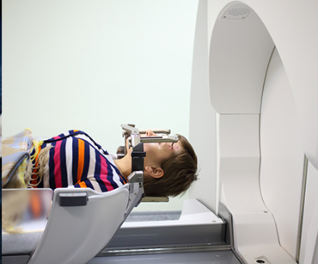
The initial steps in the LINAC procedure are similar to those in the Gamma Knife method. Doctors attach a stereotactic frame to the patient’s head with pins, then use CT or a CAT scan and MRI to determine the precise location of the tumor in the patient’s brain. Lesions up to 3.5 centimeters in diameter can be treated with the LINAC procedure. If the patient has an AVM, an angiogram and a CT scan are performed with the frame attached. Computer-guided dosimetry is specified to match the lesion. A cone that approximates the size of the lesion is placed in the collimator of the linear accelerator. The patient is placed lying on his or her back on the treatment couch of the linear accelerator. The head is secured to prevent movement during treatment. Radiation is targeted at the lesion from different directions called arcs. The arc delivers a predetermined amount of radiation, and then the treatment couch is rotated along with the collimator housing the cone. This sequence continues until the therapy is complete. The number of arcs used varies from at least four to six, and the treatment takes approximately 30 minutes. Some devices, such as the CyberKnife, do not use frames, but hold the head in place using masks instead.
Multiple manufacturers make this type of machine, which have brand names such as Peacock®, X-Knife®, CyberKnife®, Clinac®, etc.
Benefits
This technology makes it possible for neurosurgeons to reach the deepest recesses of the brain and correct disorders not treatable with conventional surgery. Since there is no incision, surgical risks such as infection are not an issue, and there is little discomfort. Adult patients may be lightly sedated but are awake throughout the procedure. Hospitalization is short and at most, requires an overnight stay. The majority of patients are treated on an outpatient basis. As a result, patients experience less discomfort and have much shorter recovery periods than having undergone conventional surgery.
Recovery
Following stereotactic radiosurgery, bandages are placed over the pin sites from the stereotactic frame, and the bandages should be removed the following day. Patients may be observed for a specified time after the treatment before they go home, or they may be kept in the hospital overnight for observation. Some people experience minimal tenderness around the pin sites. Occasionally, swelling also may occur around the pin sites. Most patients can return to their usual activities the following day if swelling is not bothersome.
Follow-up
Customarily, the neurosurgeon will want to see the patient in the office about one month after the procedure. A neurological examination will be performed. Often, a diagnostic test such as a CT scan or MRI will be performed about six months after the procedure to check on the status of the radiated area. These changes may require from one to three years to take effect.