Cerebral Aneurysms
Cerebral Aneurysms
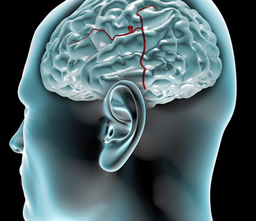
A cerebral (brain or intracranial) aneurysm is an area where a blood vessel in the brain weakens, resulting in a bulging or ballooning out of part of the vessel wall. Usually, aneurysms develop at the point where a blood vessel branches, because the “fork” is structurally more vulnerable. The disorder may result from congenital defects or from other conditions such as high blood pressure, atherosclerosis (the build-up of fatty deposits in the arteries), and less commonly, head trauma or infection.
Aneurysms are usually found at the base of the brain just inside the skull, in an area called the subarachnoid space. In fact, 90 percent of subarachnoid hemorrhages (SAHs) are attributed to ruptured cerebral aneurysms and the two terms are often used synonymously.
Aneurysm Complications Aneurysms range in size, from quite small – about 1/8 inch – to nearly one inch. Aneurysms larger than one inch are called giant aneurysms, pose a particularly high risk, and are difficult to treat.
- Saccular or “berry” aneurysms, which are shaped like a small sac, are the most common type and often require intervention.
- Fusiform aneurysms are diffuse bulges of the vessel wall involving most of the entire circumference of the blood vessel itself. They may rupture or cause stroke-like symptoms when they compress the surrounding brain.
- Mycotic aneurysms are rare and result from an infection. The infection damages and weakens the blood vessel, thereby increasing the associated risk of rupture. This infection occurs most commonly as a complication of subacute bacterial endocarditis (a heart infection). Traumatic aneurysms occur when a cerebral blood vessel incurs trauma from a head injury. The damaged blood vessel weakens at the site of injury which may cause it to rupture.
Prevalence and Incidence
- Worldwide statistics vary greatly depending on the country and demographics, ranging from as low as 5.1 cases per 100,000 persons to as high as 19.6 cases per 100,000 persons, based on age-adjusted incidence studies.
- About 40 to 50 percent of patients with ruptured aneurysms survive; 20 percent of these patients will have no permanent physical deficits.
- Re-hemorrhage occurs in about 20 percent of cases within the first 14 days after the initial rupture.
- Aneurysms occur in all age groups, but the incidence increases steadily for individuals ages 25 and older.
- Aneurysms are most prevalent in people ages 50 to 60.
- Aneurysms are about three times more prevalent in women.
- The presence of multiple aneurysms or a family history of aneurysms increases risk.
Risk Factors
- Hypertension (high blood pressure)
- Cigarette smoking/nicotine use
- Diabetes
- Excessive alcohol consumption
- Congenital (genetic) predisposition
- Injury or trauma to blood vessels
- Complication from some types of blood infections
Warning Signs/Symptoms
People who suffer a ruptured brain aneurysm may have warning signs, including:
- Localized migraine-like headache
- Nausea and vomiting
- Stiff neck
- Blurred or double vision
- Sensitivity to light (photophobia)
- Loss of sensation
Many people with unruptured brain aneurysms have no symptoms. Others might experience some or all of the following symptoms, which may be possible signs of an aneurysm:
- Cranial nerve palsy
- Dilated pupils
- Double vision
- Pain above and behind eye
- Localized headache
- Progressive weakness or numbness
Diagnostic Testing
The outcome for patients treated before a ruptured aneurysm is much better than for those treated after, so the need for adequate evaluation of patients suspected of having a cerebral aneurysm is very important.
Cerebral Angiography (also called cerebral angiogram, carotid/vertebral angiogram)
An angiogram provides accurate information that cannot be obtained from other tests. It can help detect the location and size of aneurysms and vascular malformations.
Because the test is invasive, cerebral angiography carries a measurable though small (0.5 percent) risk of stroke. Cerebral angiography is considered the gold standard test for evaluation of the blood vessels of the central nervous system.
Computed Tomographic Angiography (CTA)
CTA is a radiological test that combines the technology of a conventional CT scan with that of traditional angiography to create detailed images of the blood vessels in the brain in a noninvasive manner. This is performed by injecting a volume of contrast agent, similar to that used in the conventional angiogram, through a vein in the patient’s arm. Advantages of CTA include:
- Provides accurate identification of the exact size, location and configuration of the lesion within minutes of SAH diagnosis
- Patients do not need to be transferred to an angiographic suite
- No additional personnel are required
- There is no risk of ischemic stroke
Although in most cases CTA will provide important and accurate diagnostic information, it is less accurate and less sensitive than cerebral angiography. This fact is particularly important for small, blister-like aneurysms, or dissections (tears) of blood vessels causing SAH.
Computed Tomography (CT or CAT scan): A diagnostic image created after a computer reads x-rays. In some cases, a medication will be injected through a vein to help highlight brain structures. Bone, blood and brain tissue have very different densities and can easily be distinguished on a CT scan. A CT scan is a useful diagnostic test for hemorrhagic strokes because blood can easily be seen. CTA is the preferred diagnostic method if an aneurysm is suspected.
Magnetic Resonance Angiogram (MRA): This is a noninvasive study which is conducted in a magnetic resonance imager. The magnetic images are assembled by a computer to provide an image of the arteries in the head and neck. The MRA shows the actual blood vessels and can help detect blockage and aneurysms. MRA does not provide more information about a cerebral aneurysm than a CTA of good quality, and takes much longer to perform (about 35 minutes). It is also contraindicated in patients with certain types of metallic implants. Its advantages over CTA are that 1) patients with iodine allergy are not excluded; 2) there is no exposure to ionizing radiation.
Aneurysm Complications
Immediate Complications
- Increased intracranial pressure, direct brain destruction
Blood can leak into the cerebrospinal fluid (CSF) or areas surrounding the brain and cause an intracranial hematoma (a blood clot). Blood can irritate, damage, or destroy nearby brain cells. This may cause problems with bodily functions or mental skills. In more serious cases, the bleeding may cause brain damage, paralysis or coma. The immediate effect of aneurysm rupture is increased intracranial pressure, with immediate death occurring in 20 to 30 percent of cases.
Delayed Complications
- Brain Swelling (Edema)
The brain, like other areas of the body, has an angry response to injury. This inflammatory response may greatly increase the mass effect of a hemorrhage, and create severe conditions of brain swelling and distortion in a delayed fashion (2 to 5 days). Because the skull is closed, even small degrees of swelling may lead to significant increases in intracranial pressure.
Delayed Cerebral Vasoconstriction (Vasospasm)
Blood leaking around the brain may cause vasospasm, a narrowing of the blood vessels, which decreases the amount of blood supplied to the brain, and may result in a stroke. Vasospasm usually develops 5 to 8 days after the initial rupture, but the patient is at risk for up to 21 days (most often 3 to 14 days after SAH, with peak vasospasm occurring between days 7 to 10). To treat vasospasm, blood pressure is often purposely elevated with medications. Currently, vasospasm is not completely understood. It is the leading cause of delayed death and neurological disability in patients initially surviving the rupture of a cerebral aneurysm.
Hydrocephalus
Blood from a ruptured aneurysm can block CSF circulation, leading to hydrocephalus, a condition in which excess CSF builds up within the ventricles (fluid-containing cavities) of the brain. This may increase pressure within the head. Fluid may also build up in the brain after surgery to repair the aneurysm. To stop fluid from building up, a drain may be placed in the ventricles, to remove excess CSF and blood that has leaked. In cases where normal CSF circulation is not restored, a permanent diversion called a shunt is surgically implemented. This is necessary in about 50 percent of patients sustaining a high-grade SAH.
The combined effect of these complications creates a 30-day mortality of 50 percent for this disease, regardless of the treatment used to eliminate the aneurysm. Thus, the successful obliteration of the aneurysm is the first key consideration in the patient’s treatment.
Treatment Options
The treatment of an aneurysm, like all medical decisions, should be agreed upon by both the physician and the patient. If the case is a medical emergency, in which the ruptured aneurysm has caused the patient to lose consciousness, this discussion may take place with the patient’s nearest relative or medical decision maker. The treating physician will discuss the risks and benefits of each available treatment option. He or she will usually recommend one treatment over another, based on the patient’s individual circumstances.
While the best method of securing the aneurysm should be made on an individual basis, in general, patients with a ruptured cerebral aneurysm should be treated as soon as possible. Surgical risks and outcomes depend on whether or not the aneurysm has ruptured, the size and location of the aneurysm, and the patient’s age and overall health.
The following factors should be considered:- Risk of hemorrhage — what is the chance of the aneurysm rupturing?
- Size, shape and location
- Age and health of patient
- Family history
- Risks of treatment
- Nonsurgical Treatment
Medical therapy is an option reserved for the treatment of unruptured aneurysms in patients with significant risk factors for surgery and general anesthesia. Patients with unruptured aneurysms who have high blood pressure may be prescribed an antihypertensive (blood pressure lowering) medication, and encouraged to follow a diet and exercise program in addition to smoking cessation. These are important factors that have been shown to have a significant effect on aneurysm formation, growth, and/or rupture.
Surgery/Clipping
This method is used for both ruptured and unruptured aneurysms. Surgical repair of cerebral aneurysms is not advisable if they are located in areas of the brain that are difficult to reach surgically, or if the surgical corridor puts vital brain tissue at excessive risk. Prior to surgery, the exact location of the aneurysm is identified through cerebral angiography images. An operation to “clip” the aneurysm is performed by doing a craniotomy (opening the skull surgically), and isolating the aneurysm from the normal bloodstream. In addition, a craniectomy, a surgical procedure in which part of the skull is removed and left off temporarily, may be done to help relieve increased intracranial pressure.
One or more tiny titanium clips with spring mechanisms are applied to the base of the aneurysm, allowing it to deflate. The size and shape of the clips is selected based on the size and location of the aneurysm. Clips are permanent, remain in place, and generally provide a durable cure for the patient. Angiography is used to confirm exclusion of the aneurysm from the cerebral circulation and the preservation of normal flow of blood in the brain.
Advantages- Durability
- Direct visualization of the aneurysm (which may have very complex dimensions)
- Ability to decompress or deflate the aneurysm after clipping
- Ability to inspect other blood vessels in the area for smaller aneurysms
- In cases of SAH, the advantage of enabling evacuation of some of the clotted blood surrounding the aneurysm and the base of the brain
- Better option for convoluted arterial systems
- Better option for wide-neck aneurysms or those without defined openings
Disadvantages- Invasive surgery
- Infection
- Potential complications associated with opening the cranium
- Possible damage to other structures while clipping the aneurysm
Endovascular Surgery/Coil Embolization
Procedure
In endovascular microcoil embolization, a needle is placed into the femoral artery of the leg, and a small catheter is inserted. Utilizing x-ray guidance, the catheter is advanced through the body’s arterial system to one of the four blood vessels that feed the brain. A smaller micro catheter is fed into the aneurysm, and once properly positioned, a thin wire filament or “coil” is advanced into the aneurysm. The flexible, platinum coil is designed to conform to the shape of the aneurysm. Additional coils are advanced into the aneurysm to close the aneurysm from the inside. This prevents flow of blood into the aneurysm by causing a clot to form on the inside.
Balloon-assisted coiling uses a tiny balloon catheter to help hold the coil in place. Although this has been shown in several studies to increase risks, ongoing innovations in this relatively new technology has helped improve its efficacy. Combination stent and coiling utilizes a small flexible cylindrical mesh tube that provides a scaffold for the coiling. Intracranial stenting and other innovations are quite new, and endovascular technology is in a constant state of development. These adjuncts allow coiling to be considered for cerebral aneurysms that may not have an ideal shape for conventional coiling.
Advantages
- Minimally invasive approach does not require opening the cranium
- Provides a better outcome in older and sicker patients
- Shorter anesthesia time
- Safer option for aneurysms that are difficult to reach
- Shorter hospital stay for unruptured aneurysms – generally one-two days
- Quicker return to work – generally one to two weeks (unruptured aneurysms)
Disadvantages
- Lower chance of immediate and complete aneurysm occlusion
- Patient’s anatomy may preclude, or negate the safety of coiling because of convoluted vascular system
- Aneurysm shape should ideally consist of a relatively narrow neck and wider dome (dome to neck ratio of 2:1)
- Higher chance of aneurysm recurrence
- Slight bruising/discomfort at the catheter leg site
- Longer follow-up required to ensure treatment is durable
Post Treatment
For a patient with a ruptured cerebral aneurysm, surgical elimination of the aneurysm is only the beginning. Intensive care recovery for the next 10 to 14 days is the rule, during which time a multitude of complications related to SAH can and do occur. At some time during that period (often immediately upon completion of surgery), cerebral angiography or a substitute study is done to document that the aneurysm has been eliminated. The first 2 to 5 days after SAH represent the greatest threat of brain swelling; at which time special measures (both medical and surgical) are used to diminish the effect of swelling on intracranial pressure. Near the end of this initial period, the risk period for delayed cerebral vasospasm begins, and lasts the better part of the next 14 days. Intercurrent infections such as pneumonia are common, and hydrocephalus may develop.
Outcome
Outcome and recovery after aneurysm rupture is variable. In general, those patients who presented in good neurological condition tend to have better recoveries. Complete cure after aneurysmal SAH is achievable, and should be sought whenever possible, but recovery is often measured in months to years, rather than days to weeks. Prolonged rehabilitation, which includes physical therapy, occupational and cognitive therapy, and speech therapy, is a very important part of recovery from hemorrhagic stroke (aneurysm), just as it is for patients surviving ischemic stroke. The brain has excellent capabilities for recovery, but a long timeline. A ruptured cerebral aneurysm (aneurysmal SAH) remains among the deadliest of afflictions and carries serious, if treatable consequences. Some of these are:
- Behavioral changes
- Cognitive difficulties and short-term memory
- Decreased concentration
- Depression
- Drowsiness and fatigue
- Fine motor control impairments
- Headache
- Loss of balance or coordination
- Speech problems
- Visual disturbances
- Delayed hydrocephalus
For patients with unruptured aneurysm and uncomplicated surgery, recovery is usually complete, and relatively rapid. This does not negate the seriousness of the surgery, whether it is microsurgical or endovascular, and the recovery period for patients to return to work is often 4 to 6 weeks, despite excellent immediate outcomes and the absence of complications.